Anatomical Sciences
Research
Characterization the mechanical properties of the deep fasciae.
As regard the investigation of the mechanical behavior of the deep fasciae, the actual knowledge are very limited on the experimental side and even more on the side of the numerical modeling. The human fascial system is often quoted as the source of numerous dysfunctions, but its histological structure and biomechanical behavior are scarcely know. Usually the scientists use the data coming from the experimental tests regarding the tendons, but really the recent anatomical researches have highlighted that the fasciae present different and specific characteristics. The better knowledge of the histology of the fasciae and their mechanical behavior will permit to analyze in a scientific manner the role of the manual therapies and physiotherapy instrumentations in the care of myofascial diseases.
The overall objective of the research is to analyze the mechanical behavior of the deep fasciae by means of an integrated anatomical-histological and biomechanical investigation and to develop a specific curve of stress-strain behavior. This is a fundamental step to understand the response of the fascia to stress, overuse and immobility. Ad-hoc constitutive models based on a correct interpretation of the structural and micro-structural conformation of the fasciae will developed. In particular, it will be possible to reach a rational comprehension of the properties of anisotropy, non-linear elastic and visco-elastic behavior of the fascial tissue.
The functional response of the deep fascia to different mechanical conditions.
This could help in the comprehension of the alterations of the mechanical behavior of deep fascia that could be responsible of some myofascial pathologies, and that the manual and physical therapies can care thanks to variation of temperature, or stretch, or pressure.
The results of the mechanical experimental analyses and of the constitutive and numerical modeling will be adopted to discuss the correlations between micro-structural and macro-structural conformation of the deep fasciae of the limbs and their functional behavior. In particular it is expected that the properties of anisotropy, stiffness and visco-elastic behavior can be the relevant aspects for the discussion about the functional response of the deep fascia. The model parameters that minimize the difference between experimental and numerical stress-strain curves will be calculated. The comparison of experimental and numerical curves will be the verification of the capability of the constitutive models in describing in a suitable way the elastic and anisotropic behavior of the samples of deep fascia.
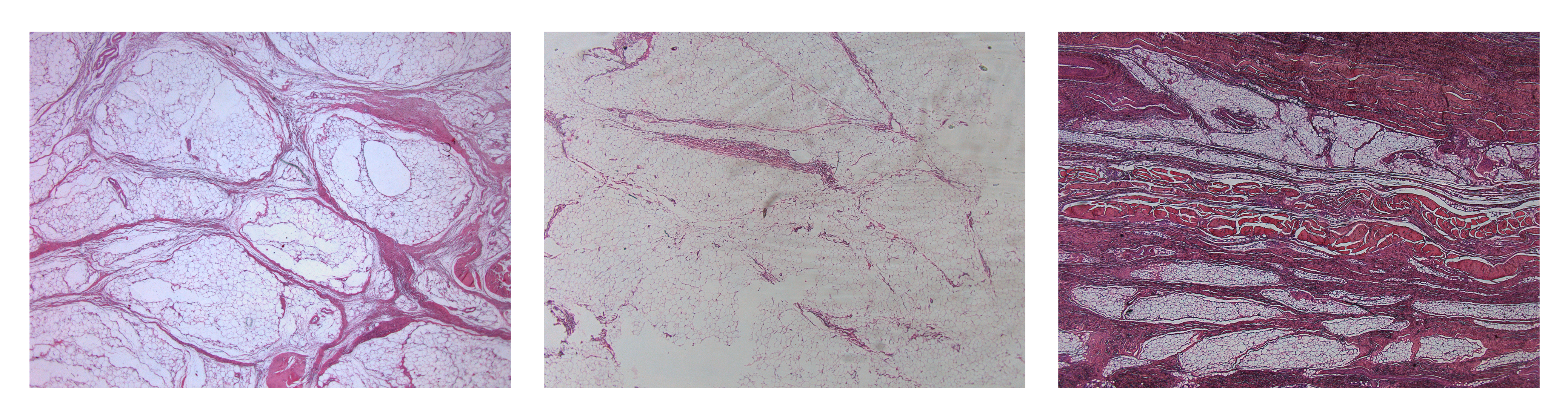
Other activities concern the histological and immunohistochemical analysis of the deep fasciae and mechanical tests.
In details, Dr Stecco’s staff takes samples of the deep fascia from different subjects both health than affected by different pathologies. Each sample is analyzed with different histological stains (HE, azan-Mallory for the collagen fibres, van Gieson for the elastic fibres and specific stains for the GAG), immunohistochemical stains (antiS100 for the nerve fibres, antiSMA for the smooth muscle, anti-sarcomeric antibody for the striated muscular fibres, antibodies for the collagen typization). Electron microscope analysis is used to study the components of the extracellular matrix, its variations between individuals, and in different ambient settings. All these instrumentations are available at the Anatomy Institute.
Mechanical tests.
The testing machines are adapted in order to develop the tests in conditions that are very similar to the physiological state. The tests aimed to the characterization of the viscoelastic behavior of the fasciae, on the evaluation of creep phenomena (change of strain of the tissue subjected to constant stress state) and relaxation (change of stress of the tissue subjected to constant strain state). The tensile tests are carried out along different directions, on the basis of the spatial configuration of the collagen fibers in the sample, to define the anisotropic characteristics of the fasciae.
Characterization the mechanical properties of the adipose tissues.
As regard the investigation of the mechanical behavior of the adipose tissues, the actual knowledge are very limited on the experimental side and even more on the side of the numerical modeling. The human adipose system is recently quoted in osteoarthritis, but its histological structure and biomechanical behavior are scarcely know.
The overall objective of the research is to analyze the mechanical behavior of adipose tissue in specific region of the body such as knee and foot, by means of an integrated anatomical-histological and biomechanical investigation and to develop a specific curve of stress-strain behavior. This is a fundamental step to understand the response of the adipose tissue also in pathogy. Ad-hoc constitutive models based on a correct interpretation of the structural and micro-structural conformation of the adipose tissue will developed.
The aim of the research activity, in collaboration with other CMBM groups, is to analyse the correlation between histological configuration and mechanical properties of different adipose tissues. Histological and ultrastructural methods are exploited to analyse the microscopic anatomies. Then, computational models are developed to evaluate the mechanical functionality. The procedure adopted allows also the investigation of tissues affected by different pathologies, considering histomorphometric and mechanical changes. This investigation is correlated with the functional response of different articular districts with regard to load bearing capabilities and movement analysis.

Educational Program
The Institute of Human Anatomy, established at University of Padova (Italy), develops advanced research and educational activities in the area of basic and advanced Human Anatomy. The researches in microscopic and clinical anatomy, neurophysiology are performed on international basis and properly documented. State-of-the-art structures of the Institute include a recently renovated dissecting room, and laboratories of cell and molecular biology, cell and tissue imaging, light and electron microscopy, radiological anatomy and clinical anatomy. At the present, the permanent staff members are 8 professors and 8 technicians, each one with specific qualifications and research activities. The Institute of Human Anatomy collaborates with numerous other international centers of Anatomy.